The most important cold laser setting isn't the device name. It's the relationship between wavelength, tissue depth, power density, energy density, treatment time, and the clinical target. A protocol that helps one anatomical site can be poorly matched to another, even when the same handpiece is used. That's why modern photobiomodulation practice treats cold laser therapy as a prescription, not as a universal machine preset.
Cold laser therapy, also called low-level laser therapy, or LLLT, has a documented origin in modern biomedical use in 1967, when Endre Mester made observations at Semmelweis Medical University in Hungary that helped establish the field of photobiomodulation (World Association for Photobiomodulation Therapy recommendations). The clinical question has since shifted from whether low-intensity light can influence tissue to which parameters should be selected for a particular condition.
Why Cold Laser Therapy Protocols Depend on Parameters
A cold laser protocol is a prescription, not a device preset. Light dose must match the tissue being treated, the target depth, and the clinical indication. A superficial lesion and a deep muscle pain generator should not receive the same settings just because one handpiece can treat both.
The prescription is more than the wavelength
A reproducible treatment record includes wavelength, output, power density, energy density, application time, pulse repetition rate when used, treatment area, and delivery method. These variables interact with tissue depth and the condition being treated, so changing one can alter the delivered dose. Professional recommendations address this parameter-based approach in the WALT recommendations.
Consider an 808 nm unit applied over the trapezius at a modest surface dose. That nominal setting may provide too little exposure at a deeper gluteal trigger point because tissue attenuates the beam before it reaches the target. Over a thin, superficial region, the same setting may expose the relevant tissue to more energy than intended. The device label alone cannot resolve that difference.
Practical rule: Identify the deepest intended target first. Then calculate the dose expected to reach it. Do not select a preset only because the device labels it “pain.”
Why one universal setting fails
Published protocols offer starting frameworks rather than fixed recipes. WALT guidance separates dosing tables for wavelengths around 780 to 860 nm and for 904 nm, supporting indication-specific prescribing rather than one setting for every musculoskeletal complaint.
Before accepting a protocol, verify:
- What tissue is being targeted?
- How deep is the target?
- Is the dose recorded per point or per square centimetre?
- Does the probe's spot size change the calculation?
- Has the stated output been verified at the applicator?
These checks expose practical differences between jaw pain, superficial tendon symptoms, and deeper muscle targets. They also clarify whether a reported dose can be reproduced in clinic. If the chart omits treatment area, delivery method, or output verification, another clinician may be unable to reconstruct the prescription or explain its selection.
Core Dosing Terms and Acronyms
Cold laser therapy protocols become easier to audit once every parameter has a defined meaning. The terms below describe different parts of the same dose calculation, and confusing them can turn an apparently precise prescription into an unreliable one.
The parameter vocabulary
- Wavelength: Measured in nanometres, or nm. It identifies the light band and helps determine tissue interaction and penetration.
- Power output: Measured in milliwatts or watts. This is the rate at which the device emits energy.
- Irradiance or power density: Measured in mW/cm². It describes power distributed across the treatment area.
- Fluence or energy density: Measured in J/cm². It describes energy delivered across a defined area.
- Treatment time: Measured in seconds. It determines how long the applicator remains over a point or area.
- Frequency: Measured in hertz, or Hz, for pulsed modes. It describes the pulse repetition rate.
Average power and peak power aren't interchangeable. In pulsed delivery, the duty cycle determines how much energy is emitted over time, so a device with a high peak output may deliver a lower average dose than the peak figure suggests.
The field also uses a biphasic dose-response model. In practical terms, both underdosing and overdosing can blunt the intended response, which is why increasing intensity without checking total energy isn't a reliable correction.
Acronyms clinicians should share
LLLT means low-level laser therapy. PBM means photobiomodulation. ATP refers to adenosine triphosphate, COX to cytochrome c oxidase, NIR to near-infrared, and WALT to the World Association for Laser Therapy.
A basic calculation provides a useful audit. A probe delivering 50 mW for 120 seconds delivers 6 J, because 0.05 watts multiplied by 120 seconds equals 6 joules. That calculation gives total energy, not energy density, unless the beam area is also known.
Wavelength Ranges and Depth of Penetration
Wavelength selection should follow the target tissue, not the diagnosis label alone. Technical guidance describes commonly used therapeutic wavelengths across roughly 600 to 1000 nm, while professional protocols distinguish between visible red wavelengths for superficial targets and near-infrared wavelengths for deeper structures (technical review in Current Pain and Headache Reports).
Visible red light, generally around 600 to 700 nm, is commonly selected for superficial lesions and surface-level tissue work. Near-infrared bands around 780 to 860 nm are frequently used when the intended target lies deeper, while 904 nm has its own pulsed protocol guidance. Above the near-infrared range, tissue absorption characteristics change, so a higher numerical wavelength isn't automatically a better deep-tissue choice.
Match the band to the clinical target
The table below is a practical orientation rather than a substitute for a condition-specific prescription. Penetration varies with tissue composition, beam geometry, contact method, and dose.
| Wavelength Band | Representative Diodes | Estimated Penetration | Typical Indications |
|---|---|---|---|
| 600 to 700 nm | Red-light diodes | More superficial | Surface lesions, mucosal areas, superficial tissue |
| 780 to 860 nm | Near-infrared diodes | Deeper soft tissue targets | Joint capsules, nerves, muscle trigger points |
| 904 nm | Pulsed diode or superluminous diode systems | Deeper peri-articular targets | Indication-specific pain and inflammatory protocols |
Chromophore absorption also matters. Cytochrome c oxidase is discussed in photobiomodulation literature as a relevant absorber in red and near-infrared ranges, but wavelength alone doesn't determine treatment success. Superficial tissue layers attenuate the incident beam, so the selected band should correspond to the deepest intended target with an appropriate clinical margin.
A useful patient-facing overview of the mechanism appears in how cold laser therapy works. For clinicians, however, the important takeaway is operational: document the target depth and explain why the selected wavelength is appropriate for that depth.
Energy Density, Power Density, and Treatment Time
Dose calculation must match the probe's output, spot size, and intended tissue target. Energy density, power density, and treatment time describe related but different variables. Energy density is the delivered energy over an area, power density is the delivery rate over that area, and treatment time is the duration required to reach the prescribed energy.
The basic equation is:
Energy in joules = power in watts × time in seconds
For a prescription written in J/cm², calculate the effective treatment area before setting the timer. A contact probe may alter the beam's effective area, so a calculation based on a non-contact aperture may not apply to contact delivery. Confirm the manufacturer's spot size and document whether the probe is stationary, moved across the target, or applied at multiple points.
Worked calculations
For a superficial example, the prescription is 4 J/cm², the probe output is 50 mW, and the treatment area is 1 cm². Convert 50 mW to 0.05 W. Delivering 4 J then requires 80 seconds per point.
For a deeper example, the prescription is 8 J/cm² and the probe output is 500 mW. At 0.5 W over 1 cm², the required dwell time is 16 seconds per point. Higher output shortens the calculated dwell time, but it does not establish that the dose suits the target or indication.
| Calculation example | Target Energy Density (J/cm²) | Probe Power (mW) | Power Density (mW/cm²) | Dwell Time per Point (sec) |
|---|---|---|---|---|
| Superficial point | 4 | 50 | 50, assuming 1 cm² | 80 |
| Deeper joint-capsule point | 8 | 500 | 500, assuming 1 cm² | 16 |
Use the equation in both directions. To calculate energy, multiply watts by seconds. To calculate time, divide the required joules by watts. If the prescription is stated per square centimetre, multiply the desired J/cm² by the treated area to obtain total joules before calculating dwell time.
The guideline summary in PMC can support a protocol review, while the WALT 904 nm dosage recommendations provide a separate reference for 904 nm dosing. Treat both as guidance for checking a prescription, not as a replacement for indication, depth, probe geometry, and treatment-area documentation.
Dose is not wattage. Increasing power density may reduce dwell time, but the prescribed energy and target depth still need to agree.
Condition-Specific Protocol Tables
Cold laser protocols are prescriptions matched to tissue depth and indication, not diagnosis labels alone. A superficial tendon, deep joint capsule, myofascial trigger point, and neuropathic pain site may all present as pain, yet require different wavelengths, beam geometry, and energy delivery.
Reported musculoskeletal protocols span approximately 600 to 1000 nm in wavelength, device outputs from 1 to 50,000 mW, and energy densities around 1 to 9 J/cm². WALT-linked guidance is organized by wavelength and indication, while the PMC technical and clinical review summarizes the broader technical and clinical variation. Use the table as a prescription-checking framework, not a universal order set.
| Indication | Tissue Depth Category | Wavelength (nm) | Energy Density (J/cm²) | Frequency | Total Sessions |
|---|---|---|---|---|---|
| Superficial tendinopathy | Superficial | 600 to 860 | 1 to 8 | Daily to several times weekly, depending on protocol | Several sessions, with reassessment |
| Myofascial trigger point | Superficial to moderate | 780 to 860 | 1 to 8, reported range | Several times weekly in reported protocols | Several sessions, insufficient consensus |
| Superficial joint osteoarthritis | Superficial to moderate | 780 to 860 | 1 to 8, reported range | Several times weekly in reported protocols | Several sessions, insufficient consensus |
| Deep joint osteoarthritis | Deep | 780 to 904 | 3 to 9 per point in reported guidance | Several times weekly, protocol-dependent | Several sessions, with reassessment |
| Post-operative wound support | Superficial | 600 to 700 | 0.5 to 8 | Daily or protocol-dependent | Until tissue response is reassessed |
| Neuropathic pain | Variable | 780 to 904 | Depth-dependent | Insufficient consensus, see text | Insufficient consensus, see text |
These ranges are not equally established. Device specifications, treatment-area size, contact technique, and reporting quality vary across studies, so a positive result in one condition should not be transferred automatically to another.
Before selecting a row, document whether the dose is prescribed per point or per square centimetre. Confirm contact versus non-contact delivery, probe spot size, and beam divergence. Those details determine the delivered fluence and make the table clinically usable rather than merely descriptive.
TMD and Orofacial Pain Protocols
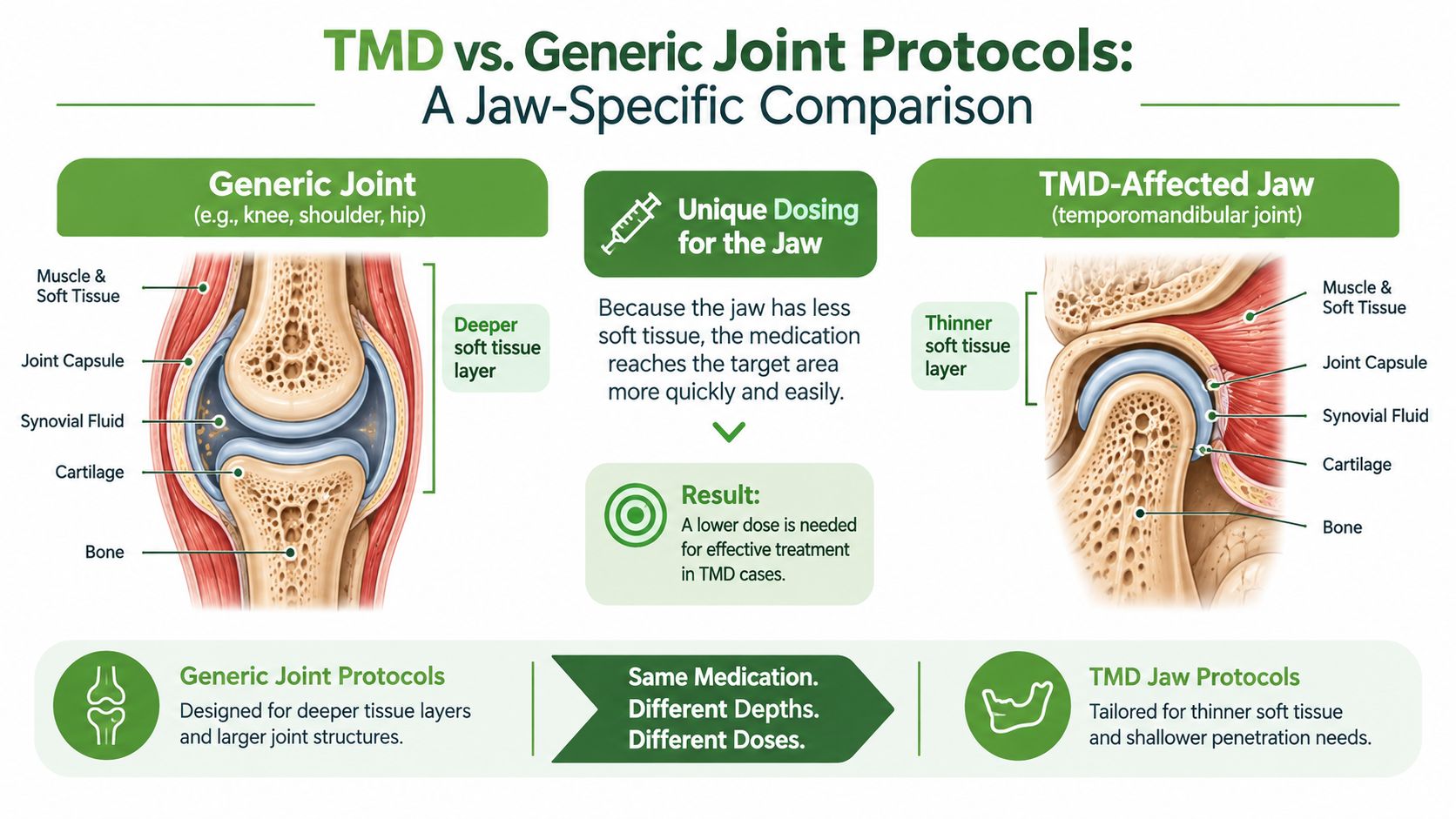
Temporomandibular disorder treatment deserves its own dosing logic. The jaw has thin overlying soft tissue in many treatment areas, and clinicians work near sensitive muscles, the joint capsule, and trigeminally mediated pain pathways. A protocol designed for a deeper shoulder or knee target shouldn't be copied onto the masseter or temporalis.
Recent synthesis supports the need for indication-specific TMD dosing. The review reports that the strongest results clustered around 810 to 940 nm and 3 to 12 J/cm², while also identifying substantial variation in wavelength, energy density, treatment duration, and reporting quality across trials (2025 TMD guideline paper). Longer treatment courses appeared more durable in that synthesis, so a one-off session shouldn't be treated as a complete therapeutic trial.
Separate the jaw targets
For extraoral masseter and temporalis work, a clinician may select a 780 to 808 nm device and prescribe a lower point dose than would be used for a deep large-joint target. Intra-oral access to the lateral pterygoid or TMJ capsule changes the optical path again, so the dose must be recalculated rather than copied from an external protocol.
The clinical plan should identify the sub-indication:
- Bruxism-related myalgia: Map the painful muscle bands and combine light treatment with load management, relaxation training, and appropriate appliance care.
- Capsulitis or joint pain: Treat the joint region with careful point selection and reassess opening, loading pain, and joint tenderness.
- Post-procedure recovery: Coordinate timing and dose with the treating dentist or surgeon rather than applying a generic jaw preset.
Pain tracking should use a structured tool, such as the graded chronic pain scale, alongside range of motion, palpation findings, headache frequency, and functional limitations. Cold laser therapy for pain relief can be considered as one component of a broader orofacial pain plan, not as a replacement for physiotherapy, occlusal management, or airway evaluation when those factors are clinically relevant.
Contraindications and Safety Screening
Cold laser therapy is non-invasive, but “cold” doesn't mean risk-free. The clinician must prevent direct ocular exposure, avoid contraindicated anatomical sites, and adjust treatment when patient factors could alter light sensitivity or tissue response.
Screen before every session
Absolute contraindications and avoidance areas include:
- Eyes: Never irradiate the eyes directly. Use wavelength-appropriate protective eyewear for everyone in the treatment field.
- Malignancy: Avoid direct treatment over known malignant tissue.
- Thyroid: Do not irradiate the thyroid region.
- Gravid uterus: Avoid treatment over the pregnant uterus.
- Active hemorrhage: Do not treat an actively bleeding area.
- Pacemakers: Avoid direct irradiation over a pacemaker unless the device manufacturer and responsible medical team provide specific clearance.
- Photosensitizing medications: Review medication changes before treatment because photosensitivity can alter risk.
Relative precautions include epilepsy, diabetes with neuropathy, children, and tattoos containing dark pigment over the treatment site. Ask about recent corticosteroid or retinoid use, new supplements, medication changes, pregnancy status, altered sensation, and prior light reactions.
Modify rather than ignore
A relative precaution calls for a revised plan, not automatic treatment or automatic abandonment. The clinician may choose a lower fluence, greater monitoring, or physical shielding, but any adjustment must be based on the specific device, tissue, and risk.
Protective eyewear should match the wavelength and the device's optical density requirements, especially for 800 to 904 nm systems. The treatment room should have appropriate warning signage, controlled access, and a closed door during emission.
Document skin inspection, eye protection verification, treatment-site screening, patient comfort, and any reaction. If the patient reports unexpected heat, visual symptoms, burning, or escalating discomfort, stop the application and reassess the device and prescription. Cold laser therapy contraindications should be reviewed alongside the device manufacturer's safety instructions and local clinical policy.
Clinical Workflow and Session Documentation
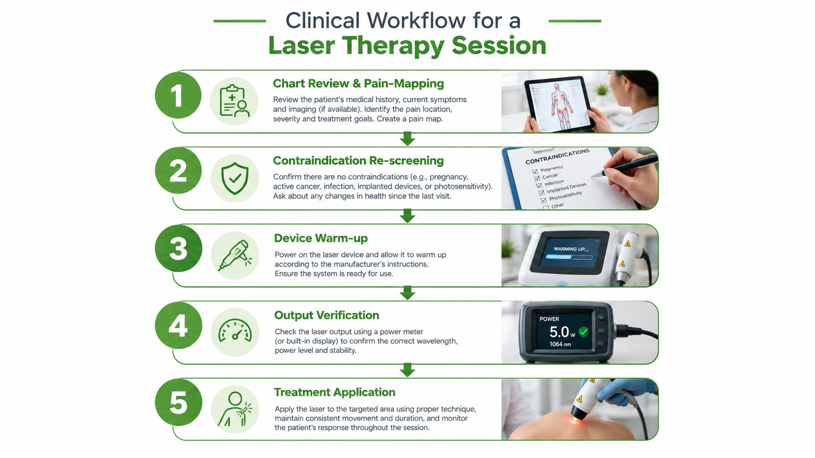
A reproducible session begins before the laser is switched on. Review the indication, map the pain, confirm the current examination findings, and ask whether anything has changed since the prior visit. New medication, pregnancy, altered sensation, a fresh procedure, or a new skin lesion can change the plan.
A practical session sequence
- Review and map: Mark the symptomatic region and relevant anatomical points on the chart or body map.
- Re-screen: Confirm contraindications, medication changes, skin condition, sensation, and patient tolerance.
- Prepare the device: Warm up the unit according to manufacturer instructions and verify output with a calibrated power meter.
- Prepare the site: Clean the skin as appropriate, establish a consistent point grid, and confirm the intended spot size.
- Apply and reassess: Deliver sequential points or a documented scanning pattern, then reassess tenderness, range of motion, and comfort.
Every chart note should contain the indication, wavelength, power at the aperture, spot size, calculated energy density per point, total joules, points treated, treatment time, pulse mode, patient response, and adverse events. If the device is pulsed, include the relevant average-output information rather than relying only on peak output.
Adjustments should be deliberate. A plateau may justify a cautious fluence change after reviewing the diagnosis, target depth, and adherence to the rest of the care plan. Heat intolerance, unexpected pain, or a new adverse response is a reason to pause, inspect the equipment, and reassess the prescription.
Evaluating Published Protocols and Trial Reports
A trial abstract can sound precise while leaving the treatment impossible to reproduce. Appraise the example: “Infrared laser therapy was applied to the painful region for several minutes at a specified power, across multiple sessions.” That statement identifies a general output and schedule, but it does not show whether the beam was stationary or scanned, how large the spot was, or how the dose was distributed.
Ask whether the reported joules refer to each point, each square centimetre, or the entire session. If the authors provide power and treatment time, total energy can be calculated from watts multiplied by seconds. If they report joules without spot size, energy density remains uncertain. A dose stated per point may represent very different exposure when the beam area changes.
A protocol isn't transparent just because it contains a wavelength.
The appraisal should also test clinical relevance. Compare the target tissue depth, application method, and stated dose with WALT tables and independent reviews, including the guideline material cited earlier. Manufacturer brochures describe device capabilities, but they do not establish condition-specific efficacy.
Look for design problems that alter interpretation: an undisclosed device class, no credible sham control when blinding is feasible, or one treatment point for a condition involving several painful structures. Check whether the authors explain the diagnosis, anatomical targets, treatment schedule, and completed sessions. A trial can remain informative, but conclusions deserve caution when the prescription cannot be reconstructed or the reported dose appears far outside the relevant therapeutic range.
Quick Reference Protocol Card
A useful protocol card should do more than hold blanks. It should show how the prescription matches the target tissue, indication, and measured dose. Verify the device output before treatment, then record the parameters in terms another clinician can reproduce.
Completed example, TMD masseter point
- Indication and target: Myofascial TMD, right masseter
- Wavelength: 808 nm
- Output power at aperture: 100 mW
- Spot size: 1 cm²
- Energy density per point: 4 J/cm²
- Total dose at this point: 4 J
- Pulse mode: Continuous
- Application: Contact, stationary placement over the marked tender point
- Treatment time: 40 seconds
The arithmetic is explicit: 100 mW equals 0.1 W, and 4 J divided by 0.1 W gives 40 seconds. If several masseter points are treated, record the number of points and multiply the per-point dose to obtain the session total. Jaw pain requires this point-by-point clarity because a small facial target should not automatically receive the same exposure used for a larger or deeper musculoskeletal region.
Minimal recording fields
- Side and landmark: ____________________
- Point category: Muscle, joint, tendon, nerve-related, or wound
- Wavelength and output: ____________________
- Spot size and dose: ______ cm², ______ J/cm²
- Energy: ______ J per point, ______ J total
- Application: Contact or non-contact, stationary or scanned
- Pulse mode and average output: ____________________
- Tolerance and reassessment date: ____________________
Record whether the stated dose applies per point, per square centimetre, or to the entire session. For a deeper target, confirm that the selected wavelength and application method suit the anatomy rather than shortening treatment by increasing power. If symptoms plateau, review the diagnosis, target selection, beam area, total dose, and concurrent rehabilitation before changing fluence.
Pain and Sleep Therapy Center provides cold laser therapy within care for TMJ and facial pain, alongside diagnostic evaluation and related pain and sleep services. Visit Pain and Sleep Therapy Center to discuss whether a parameter-matched protocol fits your symptoms and broader treatment plan.