
You may know exactly where your face hurts, but that doesn't mean you know where the pain starts. A dull ache near a molar can come from a tooth, chewing muscle, jaw joint, irritated nerve, headache mechanism, or even a problem involving nasal breathing and sleep. A brief electric shock along the cheek follows a very different diagnostic path from constant pressure around the jaw, yet both can be described as “facial pain.”
A careful facial pain diagnosis isn't based on one scan or one symptom. It develops through a sequence of questions, examination findings, targeted tests, and, when necessary, collaboration between dentistry, neurology, oral medicine, ENT, pain medicine, and sleep specialists. The process can feel slow, especially when routine tests look normal, but a systematic assessment is what prevents the wrong treatment from becoming the next source of trouble.
Why Facial Pain Diagnosis Is So Challenging
A patient may arrive after months of jaw aching, headaches, and tenderness beside the ear. One clinician suspects a tooth and recommends dental treatment. Another points to the temporomandibular joint. A third suggests sinus pressure or migraine. The patient is left with several explanations, but no clear answer.
That confusion is understandable. Teeth, periodontal tissues, chewing muscles, the TMJ, cranial nerves, sinuses, and headache pathways occupy a compact area and can produce overlapping sensations. Pain can also travel. A problem in a chewing muscle may be felt near the temple, while nerve pain can seem to come from a tooth. Pain location is a clue, not a diagnosis.

Why the first explanation may fail
A dental procedure can be appropriate when examination and testing identify a diseased tooth. It won't solve pain caused by trigeminal neuralgia, a myofascial disorder, a headache condition, or persistent idiopathic facial pain. The same problem occurs in reverse, when a scan shows a joint irregularity and clinicians assume that finding explains every symptom.
Practical rule: The more a proposed treatment changes the tooth, joint, or nerve permanently, the stronger the diagnostic evidence should be.
A second opinion should therefore involve more than another quick look at the same painful area. Bring previous images, dental records, medication history, and a timeline of what happened after each treatment. Ask the clinician to explain which findings support the working diagnosis, which findings argue against it, and what alternative causes remain possible.
The International Classification of Orofacial Pain reflects this reality by organizing facial pain according to its likely source and clinical pattern. That approach replaces guesswork based on location with a differential diagnosis built from history, examination, and appropriate evidence.
The International Classification of Orofacial Pain Framework
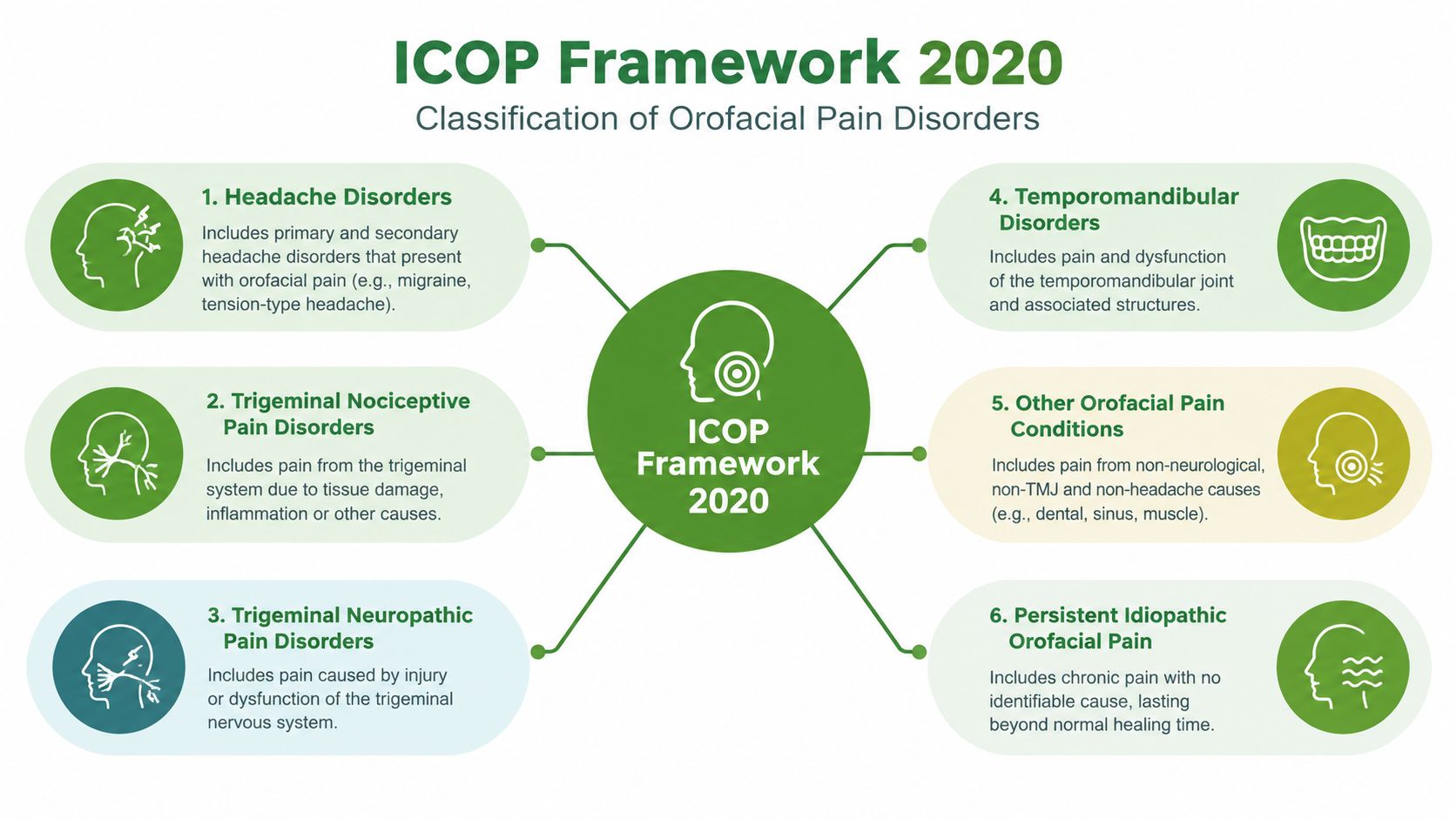
The publication of the first International Classification of Orofacial Pain, or ICOP, in 2020 marked an important step toward shared diagnostic language. ICOP recognizes that facial pain isn't one disorder and that similar symptoms may arise from different tissues or pain mechanisms.
Six categories, one clinical map
ICOP places orofacial pain into six principal categories:
- Dentoalveolar and related structural pain involves teeth, periodontal tissues, and nearby anatomical structures. The clinician looks for findings that connect the painful tooth or tissue to the symptom pattern.
- Myofascial orofacial pain arises from muscles and their connective tissues. Familiar pain reproduced during palpation or jaw function can be more informative than tenderness alone.
- Temporomandibular-joint pain concerns pain attributed to the joint itself. Joint loading, movement, sounds, restriction, and imaging may all contribute to the assessment.
- Pain caused by cranial-nerve lesions or disease includes neuropathic presentations in which the sensory pattern and neurologic findings carry substantial weight.
- Orofacial pain resembling primary headaches includes facial presentations that follow headache-related patterns rather than a dental or joint source.
- Idiopathic orofacial pain describes persistent pain that remains after appropriate evaluation has not identified a sufficient lesion or disease to explain it.
The categories don't function as labels chosen from a symptom checklist. Clinicians still need to establish onset, duration, triggers, associated features, examination findings, and whether another condition could be causing the pain. ICOP also distinguishes primary pain disorders from secondary pain caused by infection, trauma, or structural disease.
Why the framework changes treatment decisions
Without a structured framework, a painful tooth may become the default explanation only because it is easy to examine. ICOP encourages the clinician to ask whether the tooth findings truly match the pain's behavior. The same discipline applies to a joint abnormality, muscle tenderness, or an incidental radiographic finding.
For patients, this means a diagnosis may involve referral rather than immediate treatment. A dentist may involve a neurologist, an oral medicine clinician may request an ENT assessment, or a sleep-focused evaluation may become relevant when jaw symptoms occur alongside snoring, mouth breathing, or poor-quality sleep. A shared vocabulary helps those professionals describe the same problem consistently.
Patients who want a plain-language overview can also review how orofacial pain is defined and evaluated. The useful question isn't only “Where does it hurt?” It is “Which category best fits the complete pattern, and what evidence supports that conclusion?”
History and Examination The Foundation of Diagnosis
The most valuable diagnostic tool is often a well-structured conversation followed by a focused examination. Imaging can show anatomy, but it can't independently explain how pain behaves. A clinician first needs to understand the symptom in the patient's own words.
What the history should capture
Expect questions about:
- Onset and timing: Did the pain begin suddenly, gradually, after dental work, after an injury, or without an obvious event?
- Duration and rhythm: Is it continuous, episodic, or present in attacks separated by pain-free intervals?
- Location and radiation: Does it stay in one spot, cross the midline, move toward the ear, temple, eye, teeth, or neck?
- Quality: Is it aching, burning, pressure-like, stabbing, throbbing, or electric-shock-like?
- Triggers and relievers: Do chewing, speaking, brushing, touch, cold air, posture, exertion, or sleep affect it?
- Associated symptoms: Are there headaches, nausea, light sensitivity, numbness, weakness, nasal symptoms, jaw locking, clicking, fatigue, or disturbed sleep?
A pain diary can make these details easier to compare. Record the activity immediately before an episode, its character, how long it lasts, whether the jaw was moving, and what relieved it. Also list dental procedures, appliances, medications, injections, physical therapy, and the response to each.

What the examination adds
A jaw examination may measure opening and lateral movement, assess joint sounds, load the joint, and palpate the masseter, temporalis, and other muscles. The key question is whether the clinician can reproduce the patient's familiar pain, not merely find a tender spot. A muscle that recreates the usual temple ache during a specific functional test points in a different direction from a joint that produces pain during loading.
Neurologic screening evaluates facial sensation, motor function, and other cranial-nerve findings. Numbness, persistent sensory change, bilateral symptoms, or weakness may alter the urgency and referral pathway. Oral examination and dental testing remain essential, but normal dental findings should broaden the assessment rather than end it.
Breathing and sleep history also belongs in this conversation. Nasal obstruction, habitual mouth breathing, loud snoring, witnessed breathing pauses, dry mouth on waking, and morning jaw tension may reveal a contributor that isn't visible during a brief dental examination. These clues don't prove that airway or sleep breathing is causing facial pain, but they can identify an important part of the patient's overall pattern.
Diagnostic Criteria for Common Facial Pain Conditions
Different facial pain conditions require different evidence. A symptom-only approach tends to blur muscle pain, joint pain, nerve pain, and persistent idiopathic presentations. Validated protocols are useful because they combine the patient's report with reproducible examination findings.
The Diagnostic Criteria for TMD, or DC/TMD, uses a pain-focused history alongside a standardized clinical examination. In validation testing, it identified common pain-related TMD with sensitivity of at least 0.86 and specificity of at least 0.98. For myalgia, criterion validity reached 0.90 sensitivity and 0.99 specificity, while myofascial pain with referral reached 0.86 sensitivity and 0.98 specificity (DC/TMD validation findings).
Those results support a practical distinction between muscle-origin pain and intra-articular disease. Persistent locking, restricted opening, crepitus, or suspected structural joint disease may require imaging and specialist review, because clinical performance isn't equally strong for every joint disorder.
Trigeminal neuralgia follows a very different pattern. Under ICHD-3 criteria, it involves recurrent unilateral paroxysms in one or more trigeminal-nerve divisions. Attacks last from a fraction of a second to 2 minutes, feel severe and electric-shock-like, shooting, stabbing, or sharp, and are commonly triggered by normally nonpainful activities such as light touch, chewing, speaking, or brushing the teeth (ICHD-3 diagnostic criteria for trigeminal neuralgia).
A neurologic examination matters because persistent numbness, sensory loss, bilateral symptoms, or other neurologic deficits are atypical for purely classical paroxysmal disease. MRI becomes especially relevant when the pattern is unusual or secondary disease is suspected. For clinicians and patients preparing for a neurologic consultation, a focused neurology specialty resource from PatientNotes can help organize the questions and history to bring.
Persistent idiopathic facial pain presents differently again. It is uncommon but clinically meaningful. A Dutch population study reported an incidence of 4.4 cases per 100,000 person-years, with a 95% confidence interval of 3.2 to 5.9, while a German population-based study estimated lifetime prevalence at 0.03%. Other literature estimates place lifetime prevalence around 0.16% to 0.30%, with average first diagnosis in the sixth decade of life (review of persistent idiopathic facial pain). These figures must be interpreted carefully. Rarity doesn't make persistent pain insignificant, and a diagnosis requires exclusion of dental, musculoskeletal, neurologic, and structural causes.
| Condition | Key diagnostic features | Primary assessment method |
|---|---|---|
| Pain-related TMD | Familiar pain reproduced through standardized muscle or joint examination, with jaw-function findings | DC/TMD history and clinical examination, with imaging when structural disease is suspected |
| Trigeminal neuralgia | Unilateral, brief, severe electric-shock-like attacks with characteristic triggers and trigeminal distribution | Detailed temporal and sensory history, neurologic examination, and MRI when indicated |
| Persistent idiopathic facial pain | Persistent facial pain without a sufficient dental, musculoskeletal, neurologic, or structural explanation | Diagnosis of exclusion using comprehensive history, examination, and selective testing |
Imaging AI Tools and the Sleep-Airway Connection
A scan can show that a joint, sinus, tooth, or nerve has an anatomical feature. It can't automatically prove that the feature causes the patient's pain. This distinction is central when MRI, CBCT, panoramic radiographs, or thermography reveal abnormalities that are common, incidental, or unrelated to the symptom pattern.
What imaging can and can't answer
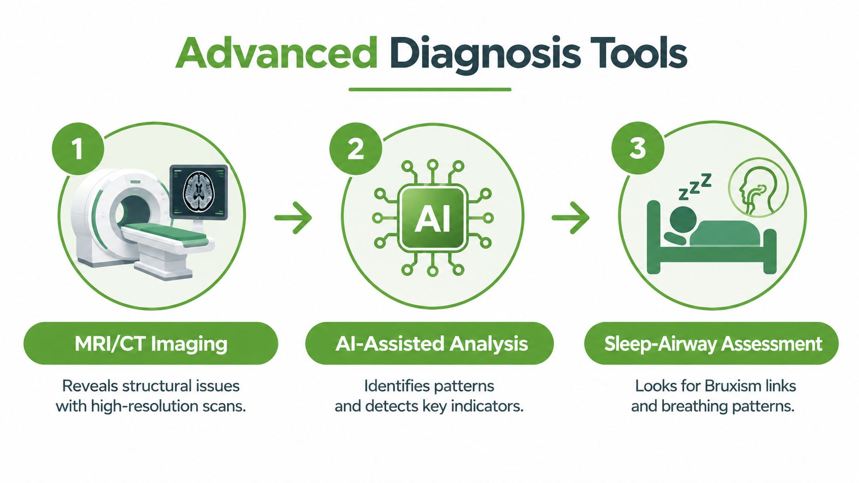
MRI may help evaluate soft tissues and joint structures. CBCT can provide detailed views of bone, teeth, joint anatomy, and the airway. Those tools are valuable when the examination raises a specific question, such as suspected structural joint disease or a possible lesion. They become less useful when ordered to find something that can be blamed for pain.
Reviews of AI-assisted interpretation report diagnostic accuracy ranging from 59% to 100%, with sensitivity from 0.76 to 0.80 and specificity from 0.63 to 0.95 (review of AI-assisted orofacial imaging). Those figures describe image classification, not whether an abnormality explains an individual person's pain, headache, chewing limitation, or muscle tenderness. AI can support pattern recognition, but it isn't an independent diagnosis and still needs clinical validation.
Why airway and sleep belong in the assessment
Facial pain evaluation should include questions about snoring, nasal breathing, witnessed pauses, restless sleep, morning headaches, dry mouth, and jaw clenching. Sleep-disordered breathing may coexist with jaw muscle overactivity or altered oral posture, while nasal obstruction can encourage mouth breathing and influence how the lips, tongue, and jaw rest. These associations are reasons to investigate, not proof of a single cause.
A sleep and airway assessment may include a detailed breathing history, nasal examination, review of sleep symptoms, and referral for appropriate sleep testing. Patients who want broader context on breathing disruptions and health risks can use that educational resource alongside professional evaluation. The point is to avoid treating a joint image while overlooking a breathing problem that may be affecting sleep and daytime function.
A practical overview of this whole-person approach is also available through dental sleep medicine. The best diagnostic pathway combines the scan with the patient's symptoms, jaw function, neurologic findings, dental status, and sleep-airway pattern.
A short visual explanation can help patients understand how these tools fit together:
How a Specialized Pain and Sleep Clinic Evaluates Facial Pain
A specialized pain and sleep clinic generally starts with the same principle as any sound facial pain diagnosis: identify the pain generator before choosing treatment. The assessment may bring together dental sleep medicine, orofacial myofunctional therapy, neurology, and pediatric oral health, depending on the patient's age and presentation.
The first visit should feel investigative rather than transactional. The clinician reviews the timeline, prior treatments, jaw function, muscle tenderness, joint mobility, oral habits, cranial-nerve findings, nasal breathing, and sleep symptoms. Technology may support the examination with targeted imaging or airway evaluation, but the results are interpreted alongside the clinical pattern.
What root-cause care may include
A patient with muscle-related jaw pain and poor oral posture may receive an exercise-based program focused on nasal breathing, tongue position, swallowing, and jaw coordination. Another patient with clear joint findings may need a different conservative plan. A person with suspected neuropathic pain may require neurologic referral rather than a dental appliance.
Non-surgical options can include a custom oral appliance when clinically appropriate, Prolotherapy, Platelet-Rich Fibrin injections, cold laser therapy, and guided exercises. These treatments aren't interchangeable, and none should be selected solely because a scan looks abnormal. The relevant choice depends on the diagnosis, risks, goals, and response to prior care.
Clinician preparation also matters. Resources on pain competencies for clinicians emphasize the value of structured assessment and communication when pain has multiple possible sources. Patients should ask what the proposed treatment is intended to change, what evidence supports the target, and what the next step will be if symptoms don't improve.
For people seeking a coordinated evaluation of jaw, facial pain, and sleep-related factors, facial pain specialists may provide a starting point for referral discussions. The appropriate clinic might still refer onward to neurology, ENT, oral medicine, or pain medicine when the findings call for expertise outside its scope.
Taking the Next Step Toward Clear Diagnosis
A clear diagnosis usually comes from better organization, not from collecting every possible test. Before an appointment, write a brief timeline of when the pain began, how it has changed, and what happened after dental work, medication, appliances, injections, or therapy. Bring copies of imaging reports and, if possible, the actual images rather than relying only on a summary.
A useful symptom record should include:
- Attack pattern: Note whether pain is continuous or occurs in brief episodes, including approximate duration.
- Pain behavior: Describe the quality, exact location, radiation, and severity without assuming the source.
- Triggers: Record chewing, speaking, touch, brushing, posture, exercise, cold air, sleep, or waking.
- Related symptoms: Include numbness, weakness, headaches, nasal obstruction, snoring, dry mouth, jaw locking, and sleep disruption.
- Treatment response: State what helped, what failed, and whether a treatment changed the pain or only caused side effects.
Ask the clinician, “What diagnoses are you considering?” and “Which finding supports each one?” It's also reasonable to ask what would make the team change direction, whether imaging is answering a specific clinical question, and whether referral to neurology, ENT, oral medicine, or pain medicine is appropriate.
Urgent assessment is warranted for new facial weakness, sudden neurologic change, difficulty speaking, a severe unfamiliar headache, high fever with marked illness, significant visual change, or rapidly developing widespread numbness. These symptoms may signal a problem outside a routine facial pain pathway.
A normal scan doesn't mean the pain is imaginary. An abnormal scan doesn't prove causation. Persistent symptoms with unrevealing dental tests deserve a broader, careful evaluation that considers nerves, muscles, joints, headache mechanisms, airway health, sleep breathing, and idiopathic pain rather than repeating an unsuccessful procedure.
Pain and Sleep Therapy Center evaluates TMJ disorders, facial pain, and sleep-related breathing concerns through individualized history-taking, head and neck examination, jaw-function assessment, technology-supported diagnostics, and sleep screening. If your symptoms overlap across dental, muscular, neurologic, and airway factors, visit Pain and Sleep Therapy Center to explore a coordinated evaluation and appropriate non-surgical care options.