You wake after a stressful week, try to yawn, and your jaw stops halfway. The opening feels smaller than usual, chewing becomes awkward, and the click you've heard for months suddenly disappears. You may wonder whether the joint has dislocated, whether the disc has torn, or whether surgery is now unavoidable.
One possibility is TMJ disc displacement without reduction, often called a closed lock. The name sounds alarming, but it describes a specific mechanical problem, not an automatic surgical emergency. Understanding what has moved, what has not, and how symptoms change over time can make treatment decisions far less confusing.
The Morning Your Jaw Won't Open
The patient described the episode clearly. After several nights of poor sleep and daytime clenching, she opened wide during a yawn and felt the jaw catch. Her mouth opened only about 25 to 30 millimeters, rather than reaching her usual range. The familiar click was gone. When she tried to move the jaw forward, it drifted toward the painful side, and the area just in front of her ear felt swollen and tender.
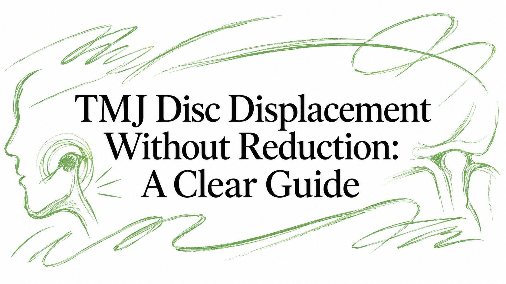
That pattern often prompts the term closed lock. In TMJ disc displacement without reduction, the disc has moved from its usual relationship with the mandibular condyle, commonly toward the front, and no longer returns to position as the jaw opens. The condyle then can't translate normally across the disc, so the person experiences restricted opening and a feeling that the jaw is physically blocked.
This differs from disc displacement with reduction. In the reducible form, the disc moves out of position when the mouth is closed but snaps back over the condyle during opening. That movement commonly produces a click or pop. In a nonreducing displacement, the click may disappear because the disc no longer moves back into place during opening.
A missing click can matter. If a long-standing click suddenly vanishes while opening becomes limited, the change deserves a clinical examination.
Not every stiff or painful jaw is a closed lock. Muscle spasm, inflammation, dental pain, trauma, arthritis, and other joint disorders can produce similar complaints. A clinician therefore looks at the timing of the change, the direction of jaw deviation, the opening pattern, joint sounds, muscle tenderness, and, when needed, imaging. You can also review practical explanations for sudden jaw locking in this guide to why the jaw locks up.
The distinction matters because disc displacement is common, while the nonreducing subtype is much less common in the general population. A systematic review estimated disc displacement overall in about 18% to 35% of people, compared with roughly 1% to 3% for DDwoR, and DDwoR accounts for approximately 5% to 15% of diagnosed TMD cases (systematic review of TMJ disc displacement). The smaller group is clinically important because it's the subtype most associated with restricted opening and persistent dysfunction.
What the Disc Does and Why It Gets Stuck
The TMJ works like a door hinge that both pivots and slides along a track. The condyle is the rounded part at the top of the lower jaw, while the fossa is the shallow area of the skull that receives it. Between them lies a biconcave fibrocartilaginous disc. It distributes force and helps the joint coordinate movement.
During a small opening, the condyle mainly rotates beneath the disc. As the mouth opens farther, the condyle and disc move forward together along the articular eminence. Normal function combines these actions, rotation followed by forward gliding, rather than relying on a simple hinge motion.

Reduction changes the sound and the movement
With a reducible displacement, the disc's posterior band sits too far forward when the mouth is closed. As opening begins, the condyle passes beneath it, and the disc returns to a more functional position for part of the motion. That transition produces the familiar click.
In DDwoR, the disc remains forward instead of moving back over the condyle. The condyle cannot pass normally over its posterior edge, so forward translation becomes limited. The jaw may drift toward the affected side because that joint is not gliding normally while the opposite joint continues to move.
Several tissue changes can contribute to this pattern. The capsule and disc-supporting ligaments may become lax, allowing the disc to shift. After displacement, the tissues behind the disc may shorten or remodel, a process often called adaptive shortening. Inflammation and fluid within the joint can increase pressure and pain, causing protective muscle tension. Later, osteoarthrotic changes may create roughness, crepitus, or additional mechanical restriction.
A visual explanation can clarify how normal movement differs from a disc that remains displaced. General information about joint displacement and dislocation care options can offer broader context, but a TMJ closed lock needs an assessment focused on the jaw joint.
The disc may stay displaced while the joint gradually develops a different working relationship. For that reason, treatment can improve pain and function without physically recapturing the disc. The appropriate goal depends on the joint's mechanics, symptoms, and how the person is functioning.
Symptoms That Point to a Closed Lock
A person with DDwoR may first notice a practical change: breakfast takes longer, biting a sandwich becomes difficult, or a dental appointment suddenly feels hard to tolerate. Some people can identify a sharp onset after yawning or biting something firm. Others develop stiffness and soreness gradually, before the limited opening becomes obvious.
Mechanical clues
The clearest mechanical finding is restricted opening. The jaw may stop at a firm endpoint, as though its path has reached a physical barrier, rather than feeling weak. An opening below roughly 35 millimeters can increase clinical suspicion, but the measurement alone cannot establish DDwoR. During protrusion or side-to-side movement, the jaw may drift toward the affected joint.
A previous reciprocal click that disappears as the jaw becomes restricted is another meaningful pattern. The change suggests that the disc is no longer moving back into position during opening. Grinding or crackling points clinicians toward a separate question, such as surface irregularity or degenerative change, so those sounds require their own assessment.
Pain and protective muscle tension
Pain often appears in front of the ear and may worsen with chewing, speaking, or assisted opening. The masseter and temporalis muscles can become tender as they brace around a painful joint. Their secondary pain may spread across the face, making the entire side feel tight.
Pain intensity does not reliably show how far forward the disc appears on MRI. Some people have imaging changes without symptoms, so clinicians compare the scan with movement, function, and examination findings. A Canadian Dental Association review described MRI studies in which disc displacement appeared in 35% of asymptomatic volunteers, supporting clinical correlation (MRI review of TMJ internal derangement).
Referred sensations
Ear fullness, tinnitus, temple headaches, and discomfort into the neck can accompany TMJ disorders, yet these symptoms do not identify DDwoR by themselves. Ear and neurologic conditions may produce similar sensations. If nighttime clenching or grinding aggravates the jaw, practical advice on home remedies for teeth grinding may support self-care, but it should not replace assessment of a newly limited opening.
The working pattern is restricted movement plus a compatible history. A forward-appearing disc on a scan, without matching symptoms and impaired function, does not by itself prove a closed lock. Each patient's treatment plan therefore depends on the combination of mechanical restriction, pain, duration, and day-to-day function.
How a Clinician Confirms the Diagnosis
Diagnosis builds in layers, combining history, movement testing, and imaging when needed. No single symptom, sound, or scan can establish a closed lock because several jaw conditions restrict opening.
History comes first
The clinician asks when the limitation began, whether it followed yawning, trauma, dental treatment, or prolonged clenching, and whether the change was sudden or gradual. A previous click that disappeared when the jaw became restricted can support the history of a disc that stopped reducing. Changes in eating, speaking, sleep, oral hygiene, and tolerance of dental care show how the problem affects daily function.
The examination measures maximum interincisal opening, the distance between the upper and lower front teeth at the widest comfortable opening. It also compares unassisted and assisted opening, lateral excursions, protrusion, deviation, joint tenderness, and muscle findings. A firm endpoint, limited forward movement, or deviation toward one side can strengthen the clinical impression, especially when these findings match the patient's history.
The examination separates similar problems
The clinician may listen for clicking, popping, or crepitus and check whether joint loading reproduces the patient's familiar pain. Muscle tenderness can contribute to symptoms, but it does not prove disc displacement. Diagnostic blocks may help identify whether pain comes mainly from the joint or from surrounding muscles when the source remains uncertain.

Imaging answers a different question
A panoramic radiograph can screen for broad dental and bony concerns. MRI is the key study for disc position and soft-tissue morphology, particularly when the clinical picture is uncertain or symptoms remain significant. CBCT is better suited to suspected bony remodeling, erosion, fracture, ankylosis, or another osseous problem.
The imaging assessment asks more than whether the disc sits forward. It examines whether the disc reduces during opening, its shape, joint fluid or inflammation, and signs of degenerative bone change. MRI findings must still be matched with symptoms, movement, and function, because an imaging abnormality may not explain the patient's limitation. A bone scan may be considered in selected cases of suspected active osseous disease, as described in this three-phase bone scan resource.
A final diagnosis therefore combines history, movement testing, pain localization, and imaging when indicated. This layered process helps distinguish a persistent mechanical restriction from muscle-related limitation, degenerative disease, or an incidental scan finding.
Treatment Options From Conservative to Surgical
Treatment should match the person, not just the scan label. Someone with pain but useful opening may need a different plan from someone with a persistent closed lock who cannot eat normally. The clinician first identifies the main target: pain control, restored motion, lower joint irritation, protection from overload, or treatment of confirmed structural disease. A displaced disc does not automatically require an operation.
The treatment ladder
Tier 1, education and rehabilitation. Initial care often combines temporary diet changes, avoidance of extreme opening, and appropriate analgesic or anti-inflammatory medication when medically safe. Physical therapy may include gentle mobilization, controlled stretching, posture work, and exercises that improve coordinated jaw movement. The aim is to restore tolerable function without repeatedly forcing a restricted joint.
Tier 2, oral appliances. A stabilization splint may reduce harmful loading in selected patients, particularly when clenching, grinding, or muscle overactivity contributes to symptoms. An anterior repositioning appliance may fit a narrower clinical pattern and requires monitoring. These appliances do different jobs. Prolonged use without review can produce unwanted bite or joint effects.
Tier 3, joint procedures. Arthrocentesis irrigates and distends the joint. It may reduce inflammatory substances and improve movement. Depending on the diagnosis, risks, and local expertise, clinicians may combine it with hyaluronic acid, corticosteroid, or platelet-rich products. Comparative evidence remains mixed. A systematic review of 20 studies involving 1,305 patients found no statistically significant difference across many conservative and surgical comparisons, although many treatments improved pain or function from baseline. Because the evidence was heterogeneous, the authors recommended starting with simple, low-risk care (systematic review of conservative and surgical TMJ treatment).
Tier 4, advanced minimally invasive care. Arthroscopy can inspect and treat selected problems inside the joint when symptoms continue despite basic care. Regenerative injections may address inflammation and function, but they should not be presented as a mechanical method for returning a displaced disc to its original position.
Tier 5, open surgery. Arthroplasty, discectomy with or without an implant, and total joint replacement are considered only for carefully selected patients with structural disease and substantial symptoms after appropriate conservative treatment.
Matching the option to the phenotype
| Treatment Tier | Best-Fit Patient | Primary Goal |
|---|---|---|
| Education, medication, diet, physical therapy | Painful joint with usable movement or early limitation | Calm symptoms and restore controlled function |
| Stabilization or selected repositioning appliance | Joint overload, clenching, or a clearly defined appliance indication | Reduce loading and support movement |
| Arthrocentesis or injection | Persistent joint pain or limitation after basic care | Improve motion and reduce intra-articular irritation |
| Arthroscopy | Refractory symptoms with a treatable intra-articular problem | Treat selected joint pathology through a minimally invasive route |
| Open surgery or replacement | Confirmed structural destruction with major ongoing disability | Reconstruct or replace the damaged joint |
People comparing approaches can review guidance on how to relieve joint pain with personalized guidance, but TMJ procedures still require individualized assessment. Evidence supports a least-invasive starting point. A 2022 systematic review of exercise and manual therapy found that exercise and manual therapy can improve outcomes, with exercise potentially improving opening more than splints, although confidence is limited by differences among studies. A broader guide to non-surgical TMJ treatment options can help patients compare nonsurgical approaches before discussing procedures with a clinician.
What Recovery Looks Like Over Time
Recovery has two separate outcomes. Symptom recovery means less pain, wider or more comfortable opening, easier chewing, and steadier function. Disc recapture means the displaced disc returns to a more typical position. These changes may occur independently, so meaningful recovery can happen even when the disc remains displaced.
Natural-history research supports measured expectations. Symptoms and movement may improve gradually rather than resolve immediately, and a displaced disc can also be present without symptoms. One longitudinal study reported clinical or spontaneous resolution in 34.1% at six months, 50.0% at 12 months, and 68.2% at 18 months (longitudinal follow-up of anterior DDwoR). A Canadian review cited successful resolution in 68% by 18 months among 44 observed patients, while emphasizing that imaging findings do not always correspond to symptoms.
A practical recovery map
Early care focuses on reducing excessive joint loading, preserving tolerable movement, controlling pain, and choosing foods that do not require wide opening. A physical therapist or clinician can set an appropriate stretching range. Forcing the jaw beyond that range may increase muscle guarding and make movement harder.
Over the following weeks and months, many patients gradually tolerate more chewing and regain confidence with speaking and oral hygiene. Progress is often uneven. A better opening one day followed by stiffness the next does not, by itself, show that the joint has deteriorated.
Long-term results vary by symptom pattern, joint condition, and response to care. A prospective cohort found that about 40% were symptom-free after 2.5 years, 33% improved, and 25% remained symptomatic or needed treatment (natural course of untreated symptomatic DDwoR). These findings support planned follow-up, especially when restricted movement affects daily activities.

The most useful long-term measure is practical function, not a perfectly normal MRI. Track whether you can open, eat, speak, sleep, and handle ordinary jaw demands without rising pain. Gradual return to activity, attention to clenching, and patient-led exercises may matter more than repeatedly checking disc position.
When It Is Time to Seek Specialty Care
A closed lock does not require surgery by itself, but it does require the right level of assessment. Evidence reviews have not identified one clear winner between conservative and surgical approaches. Many treatments improve pain or function, so clinicians usually start with lower-risk care (evidence review of DDwoR treatment).
Do not wait indefinitely if restricted opening persists, worsens, or interferes with eating, speaking, brushing, sleep, work, or dental treatment. Assessment may involve a dentist with TMJ expertise, an orofacial pain clinician, an oral and maxillofacial surgeon, and a physical therapist. Imaging can show disc position and joint structure, while the hands-on examination shows how the joint and muscles function together.
Reasons to arrange a focused evaluation
- Persistent mechanical limitation: Opening remains locked or substantially restricted despite sensible self-care.
- Progressive symptoms: Pain, stiffness, bite changes, joint noise, headaches, or ear-related sensations occur more often or become harder to control.
- Daily activity disruption: Eating, oral hygiene, speaking, or sleep repeatedly becomes difficult because of jaw symptoms.
- Unclear diagnosis: The pattern does not fit a straightforward muscle or joint problem, or earlier treatment did not address the likely cause.
Surgery deserves consideration when examination and imaging point to a meaningful structural problem, and significant pain or functional loss continues after a well-conducted conservative course. An MRI report that says “anterior disc displacement” is not, by itself, a reason for an operation. A disc that stays displaced can behave differently from a reducible disc, so treatment decisions should follow the closed-lock pattern, the joint findings, and the patient's actual limitations.

Urgent assessment is needed after facial trauma or with fever, redness, marked swelling, pus, severe unexplained pain, difficulty swallowing or breathing, rapidly worsening inability to open, suspected fracture, neurologic changes, or signs of destructive joint disease. Infection and other serious conditions can resemble a routine closed lock, and delay may complicate treatment.
If your jaw recently stopped opening normally, avoid repeatedly forcing it and do not choose surgery from an imaging report alone. Record when the change began, which movements hurt, whether a previous click disappeared, and how eating or speaking has changed. Pain and Sleep Therapy Center evaluates TMJ pain, jaw dysfunction, headaches, and related sleep or breathing concerns. Its assessment may include physical therapy, oral appliances, PRF injections, or cold laser therapy. Visit Pain and Sleep Therapy Center to request an assessment based on your symptoms and functional goals.